
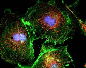
A fluorophore/fluorochrome is a type of fluorescent dye used to mark proteins, tissues, and cells with a fluorescent label for examination by fluorescence microscopy. A fluorophore works by absorbing energy of a specific wavelength region, commonly referred to as the Excitation Range, and re-emitting that energy in another specific wavelength region, commonly referred to as the Emission Range.
In general, a fluorophore will absorb energy by high frequency illumination, such as wavelengths in the ultraviolet, violet, or blue region of the spectrum, and emit energy at slightly lower frequencies, such as wavelengths in the green, red, or NIR region of the spectrum. Each fluorophore has a wavelength at which it absorbs energy most efficiently as the Peak Excitation (nm), and a corresponding wavelength at which the maximum amount of absorbed energy is re-emitted as the Peak Emission (nm). Selecting individual optical filters with the maximum amount of transmission at each of those wavelengths will ensure brilliant fluorescent images.
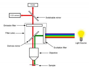
Excitation Filter
The excitation filter is placed within the illumination path of a fluorescence microscope. Its purpose is to filter out all wavelengths of the light source, except for the excitation range of the fluorophore under inspection. The Minimum Transmission % of the filter will dictate the brightness and brilliance of images. Honour Optics recommends a minimum of 40% transmission for any excitation filter, with an ideal transmission being >85%. The Bandwidth of the excitation filter should fall entirely within the excitation range of the fluorophore, with the ideal filter having a Center Wavelength (CWL) as close as possible to the peak excitation of the fluorophore in question. The Optical Density (OD) of the excitation filter will dictate the darkness of the background image, as optical density is a measure of how well a filter blocks wavelengths outside of its bandwidth. Honour Optics recommends a minimum optical density of 3.0, with an ideal optical density of 6.0. An optical filterswith the ideal combination of Center Wavelength, Minimum Transmission %, and Optical Density will provide the brightest possible images, with the deepest possible blocking, ensuring detection of the faintest emission signals.
Emission Filter
The emission filter is placed within the imaging path of a fluorescence microscope. Its purpose is to filter out the entire excitation range of the fluorophore under inspection, and to transmit the emission range of that fluorophore. The same recommendations for Minimum Transmission, Bandwidth, Optical Density, and Center Wavelength for Excitation Filters holds true for Emission Filters. Again, an emission filter with the ideal combination of Center Wavelength, Minimum Transmission %, and Optical Density will provide the brightest possible images, with the deepest possible blocking, ensuring detection of the faintest emission signals.
Dichroic Filter or Beamsplitter
The dichroic filter or beamsplitter is placed in between the excitation filter and emission filter, at a 45° angle. Its purpose is to reflect the excitation signal towards the fluorophore under inspection, and to transmit the emission signal towards the detector. An ideal dichroic filter or beamsplitter will have a sharp transition between maximum reflection and maximum transmission with a reflection % >95% for the bandwidth of the excitation filter, and a transmission of >90% for the bandwidth of the emission filter. The filter should be selected with the Intersection Wavelength (λ) of the fluorophore in mind, to ensure minimal stray-light and a maximum signal-to-noise ratio in the fluorescent image. Although selection of an appropriate dichroic filter or beamsplitter is tricky.

For fluorescence microscopy applications using multiple fluorophores, laser sources, alternate dichroic filters or beamsplitters, or applications more complex than typical fluorescence microscope setups, contact us to discuss your specifications, please e-mail to [email protected].