
What is Laser Raster Microscopy?
Laser raster microscopy, or confocal laser scanning microscopy, is an optical analysis technique for fluorescence microscopy. It is a broadly applied image analysis method with applications in biological and medical research, and utilizes a combination of a diffraction-limited laser, beam splitters, and bandpass filters, to observe the excitation of photo-fluorescence particles, or fluorescent dyes in a sample. The data from this analysis method is quantified and computerized to obtain high-quality, 3D images of samples at a microscopic level.
Instrumentation in Laser Raster Microscopy
The term raster, in laser raster microscopy, refers to the linear direction of the laser beam’s motion, which is controlled either by highly-tunable dichroic mirrors inside a fluorescence microscope, or by minutely moving the sample. Either process is capable of accurately steering the laser beam to rapidly scan the sample in a series of sweeping parallel lines. This continuous linear motion, combined with the diffraction-limited potential of the input laser makes it possible to scan miniscule sample sections and observe fluorescence from exact points on the nanoscale.
Fluorescence microscopes for laser raster microscopy require a diffraction-limited input laser beam that is directed at a robust dichroic beam splitter, which are available with varying wavelength transmissions based on the wavelength of the laser. This optical characteristic is crucial, as fluorescence signals are thousands of times weaker than the laser light used to excite a sample. Observing microscopic fluorescence through laser raster microscopy therefore demands extreme instrumental stringency for rejecting out-of-band photons from reaching the resulting photodetector. Hence the implementation of both dedicated dichroic mirrors and robust wavelength filters capable of selecting wavelengths pertaining to fluorescence emission while filtering out background noise of exorbitantly higher brightness, such as scattered light.
These bandpass filters are implemented below a keyhole passage to the photodetector, allowing for comprehensive remittance of fluorescent photons and elimination of residual light. This process, including the motion of the input beam in laser raster microscopy, can be fully computerized, with CCD or CMOS camera attachments available to replace the eye piece component of the photodetector.
Fluorescence Microscopy Products from Honour Optics
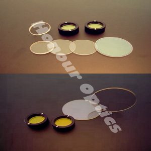
We provide a range of dichroic mirrors for laser raster microscopy, and suitable bandpass filters with ultra-hard laser-resistant coats and center wavelengths across much of the full spectrum. If you have any queries about the suitability of products from our range to your specific laser raster microscopy specifications, please do not hesitate to contact us via e-mail [email protected].