
Applications for Fluorescence Microscopy
Fluorescence microscopy is a technique used to analyze biological structures in a sample using a white lamp, and either organic or inorganic fluorophores such as dyes to excite a photo-emissive reaction, which is observed using an optical bandpass filter and a dichroic mirror. Various biochemical industries rely on fluorescence microscopy for the performance of molecular imaging to support medical diagnostics or research into cellular structures and nanomolecular activity. With this article will explore some of its immediate applications and implications of further study in the field in more detail.
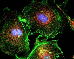
Fluorescence Microscopy for Cell Labeling
Labeling cell structures with fluorophores such as fluorescent dyes drastically increase the observable properties of biological or chemical structures. These can be modified or conjugated to exhibit specific targetability for use in various medical applications. Fluorophores with high specificity are routinely used in medical diagnostics, with increasing research finding further applications for fluorescent cell labeling.
For example: researchers have successfully used fluorescence microscopy to track and analyze viral DNA in cells at a local level and further connect that to cellular anti-viral defenses. This has potentially tremendous implications for future viral analysis and categorization.
Fluorescence Microscopy for Protein Characterization
Intra-cellular dynamic protein interactions are crucial for a range of critical biological processes, acting as catalysts for metabolic reactions or responding to stimuli. Protein interactions are typically observable using dedicated instrumentation capable of analyzing the samples at the nanometer scale. However, transient interactions of proteins display weaker signals which are typically transparent, even to fluorescence microscopy instrumentation.
The implementation of fluorescent protein biosensors is a novel approach to observing weaker protein interactions, which has found broad applications in varying fields of research. Researchers have utilized fluorescent proteins to observe intra-cellular reactions in the hopes of gaining insight into various transmission reactions, by analyzing the specific wavelengths emitted by excited cells during protein synthesis.
Such studies have tremendous potential to provide insights into how irregular translations in a cell can result in developmental problems and disease transmission – yet they also require exceptionally sensitive instrumentation and highly tunable chromatic filters to accurately assess data.
Fluorescence Microscopy with Honour Optics
We provide a range of continuously variable filters capable of examining the spectral properties of a given sample with exceptional transmission values and deep blocking in the UVA/VIS/NIR wavelength range. If you would like any more information about our products suitable for the performance of fluorescence microscopy, please contact us via email [email protected].